Ultrasound Scans in Pregnancy: What Each Trimester Involves
Ultrasound scans are a routine part of pregnancy care in Malaysia, but many parents do not realise that the purpose and focus of scans changes significantly across the three trimesters. Understanding what each scan is looking for — and why — can help you feel more prepared at every stage.
In the first trimester (weeks 1 to 13), ultrasound is primarily about confirming and dating the pregnancy. The dating scan checks that the pregnancy is inside the uterus, confirms the baby's heartbeat, and measures the baby to establish your due date. This is also when the nuchal translucency (NT) scan is performed between 11 and 14 weeks, screening for chromosomal conditions. First-trimester scans are sometimes done transvaginally for clearer images, especially early in the pregnancy.
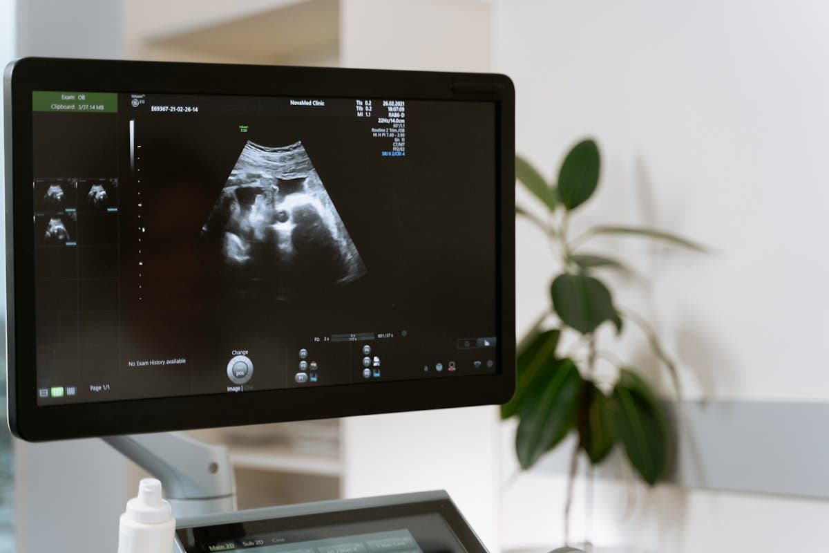
The second trimester (weeks 14 to 27) is when the most important structural assessment takes place. The detailed anomaly scan at 18 to 22 weeks examines every major organ system in the baby's body. This is the scan where the quality of the operator matters most — an MFM specialist like Dr. Kartik, with advanced fetal imaging training, is best placed to perform and interpret this examination. The second trimester is also when growth patterns begin to be tracked and when the placenta's position is assessed.
In the third trimester (weeks 28 to 40), ultrasound shifts focus to growth and wellbeing. Growth scans measure the baby's estimated weight, check the amniotic fluid volume, and assess blood flow using Doppler studies. These scans are particularly important for pregnancies with gestational diabetes, hypertension, or other risk factors. The baby's position is also checked — whether head down, breech, or transverse — which affects the delivery plan.
Doppler ultrasound, used mainly in the second and third trimesters, measures blood flow in the umbilical artery, middle cerebral artery, and uterine arteries. Abnormal Doppler results can indicate that the baby is not getting enough nutrients or oxygen, prompting closer monitoring or earlier delivery. This is a specialised assessment that benefits from MFM expertise.
If something unusual is found at any stage, additional focused scans may be arranged. Fetal echocardiography looks at the baby's heart in detail. Cervical length scans assess the risk of preterm birth. Serial growth scans track babies that are measuring small. Each of these adds information to the overall picture of your pregnancy.
Across all trimesters, Dr. Kartik Balaraman performs and interprets ultrasound scans personally at Columbia Asia Hospital Bukit Jalil. This continuity means he builds a complete picture of your pregnancy as it develops, rather than interpreting a single scan in isolation. If you would like your scans performed by an MFM specialist, you can book directly without a referral.
Further reading
- Ultrasound Scans in Pregnancy — NHS (UK)
- Doppler Ultrasound in Obstetrics — ISUOG (International Society of Ultrasound in Obstetrics and Gynecology)
Frequently Asked Questions
How many ultrasounds do you get in a normal pregnancy?
Typically three: a dating scan, an NT scan, and an anomaly scan. Additional scans depend on your individual pregnancy and any risk factors identified.
Can too many ultrasounds harm the baby?
Diagnostic ultrasound at standard clinical levels has not been shown to harm the baby. However, ultrasounds should be performed for medical reasons, not entertainment.
What is a Doppler scan?
A Doppler scan measures blood flow in the umbilical cord, the baby's brain, and the mother's uterus. It helps assess whether the baby is receiving adequate oxygen and nutrients.
Have Questions About Your Pregnancy?
Speak with Dr. Kartik Balaraman directly for personalised guidance.
Book Consultation